Nuclear medicine
Find out everything you need to know about nuclear medicine and what diseases it treats. We tell you what it is, its field of study, what its main tests involve and the different subspecialities it is usually divided into. Find the nuclear medicine unit that can help with your treatment or diagnosis and book your appointment at one of our hospitals.


Artículos de tu canal Nuclear medicine
- tu canal de salud
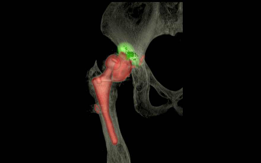
Linfogammagrafía de ganglio centinela en el cáncer de mama
What is nuclear medicine?
Nuclear medicine is a speciality that uses radioactive isotopes in a controlled manner for the treatment and diagnosis of diseases. Radiopharmaceuticals or radiolabels allow information to be obtained about organs at the molecular level, which facilitates early diagnosis and improves treatment and prognosis.
The doctors in this unit are experts in state-of-the-art technology and use it for the benefit of the patient. Contrary to what you may think, nuclear medicine techniques are not aggressive and do not require hospitalisation. Exposure to radiation is always kept to a minimum, and is similar to most radiological tests. In most cases, the patient can go back to their usual routine provided they follow minimal advice, such as staying away from children and pregnant women for a few hours.
What does nuclear medicine study/treat?
Nuclear medicine provides a functional image representing the metabolic activity of the different organs and tissues of the human body, and therefore it provides imaging of many different tissues and organs. Functional disorders generally precede morphological disorders that can be detected with other techniques, which is why nuclear medicine scans are key in ruling out major pathologies such as metastases in extension studies in oncology. But nuclear medicine is also used to make an early diagnosis of many other diseases, such as coronary heart disease or certain myocardial diseases, such as cardiac amyloidosis, Alzheimer’s or Parkinson’s disease. It also helps pinpoint the origin of certain endocrine system diseases, such as hyperparathyroidism, so that the surgeon can resort to the least invasive surgery to cure the disease. Radiodirected surgery is an example of this practice, and it involves the radiolocalisation of the sentinel lymph node, mainly in breast cancer. This allows surgeons to perform selective biopsy procedures without having to resort to larger incisions, and therefore patients benefit from a faster, safer surgery and an earlier discharge date. It is also used for the very early assessment of treatment outcomes, which are sometimes aggressive, in order to detect the need to change them if they are not effective.
Doctors can specialise in two different types of nuclear medicine with a view to providing the most suitable care to patients:
- Diagnostic imaging: use of up to 100 different substances that do not pose a health risk and do not have serious side effects. The advantage of using these elements is that they provide both morphological images, which show the shape and characteristics of an organ or tissue, and functional images, which show physiological processes.
- Therapeutic treatments: focus on certain types of thyroid cancer, prostate cancer or neuroendocrine tumours. The procedure involves the use of radioactive drugs that, after intravenous injection, bind to tumour cells and destroy them.
Techniques, procedures and diagnostic methods
There have been many technical advances in nuclear medicine in recent times, and further developments are expected in the coming years. The therapeutic techniques and diagnostic methods it uses are combined with many other specialties to detect, rule out or treat numerous diseases. The most commonly used procedures are:
- SPECT-CT scanning: a morphological CT image that allows precise localisation of the disorders detected by nuclear medicine imaging and helps understand their characteristics.
- PET-CT scanning: this test, which is one of the major advances in nuclear medicine, allows early and more reliable detection of tumours, and provides information about their extent. Combines CT and PET technology to provide 3D images of organs. It uses harmless compounds including glucose, which has a tendency to deposit in metabolically inefficient tissues like many tumours, among others.
- PET-MRI scanning: it unifies in a single process the information on molecular processes provided by the PET scan and the morphofunctional information on tissues provided by magnetic resonance imaging (MRI).
- Gamma scan: uses a gamma camera that detects radiation that has been previously injected into the patient, and shows 2D images of organs and tissues. This test provides both functional and morphological information.